Growing Tumors in Eggs to Decode Cancer
Many biological experiments involve working with live mice or watching cells develop in petri dishes.
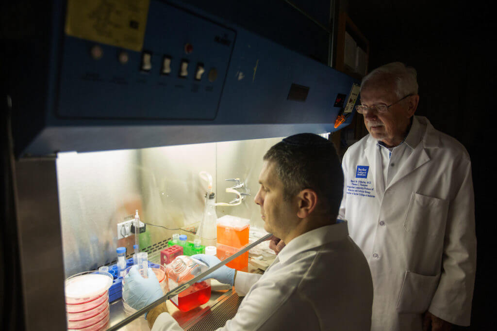
That’s why Baylor College of Medicine prostate cancer researcher David Rowley, Ph.D., was intrigued last year when he heard a colleague speak about using fertilized eggs to grow tumors.
The new take on an old idea emerged during a seminar in which Andrew Sikora, M.D., Ph.D.—an otolaryngologist (specializing in diseases of the ear, nose and throat) and researcher at Baylor—described how he performed research in his laboratory.
“Before Andrew could get off the stage, my colleague, Rebeca San Martin, and I were up there talking with him,” said Rowley, a professor in Baylor’s Dan L Duncan Comprehensive Cancer Center and leader of the college’s cancer biology program.
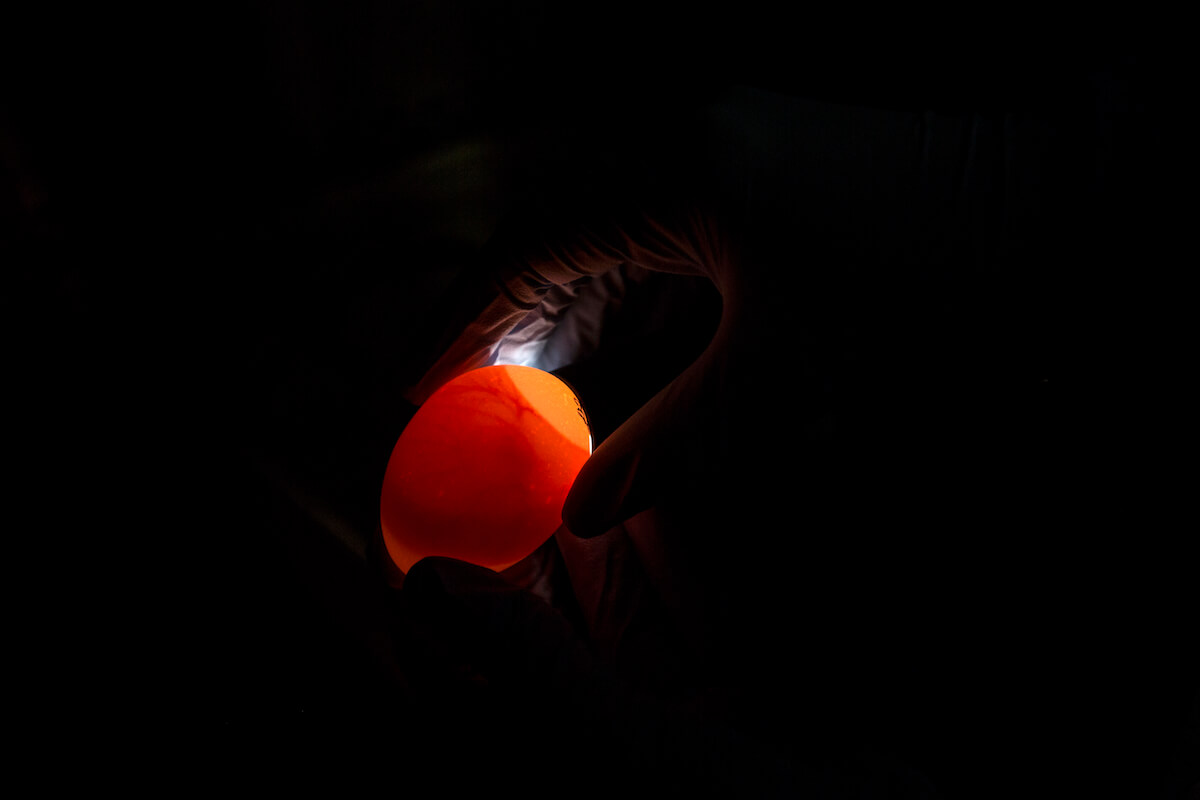
Sikora has updated the 100-year-old research technique—growing tumors in fertilized chicken eggs—for the 21st century. While studying its history, he found an article that explained the original research and other early studies.
“It’s totally old school—they had the idea to grow tumors in chicken eggs, and they actually did it and got them to grow,” Sikora said.
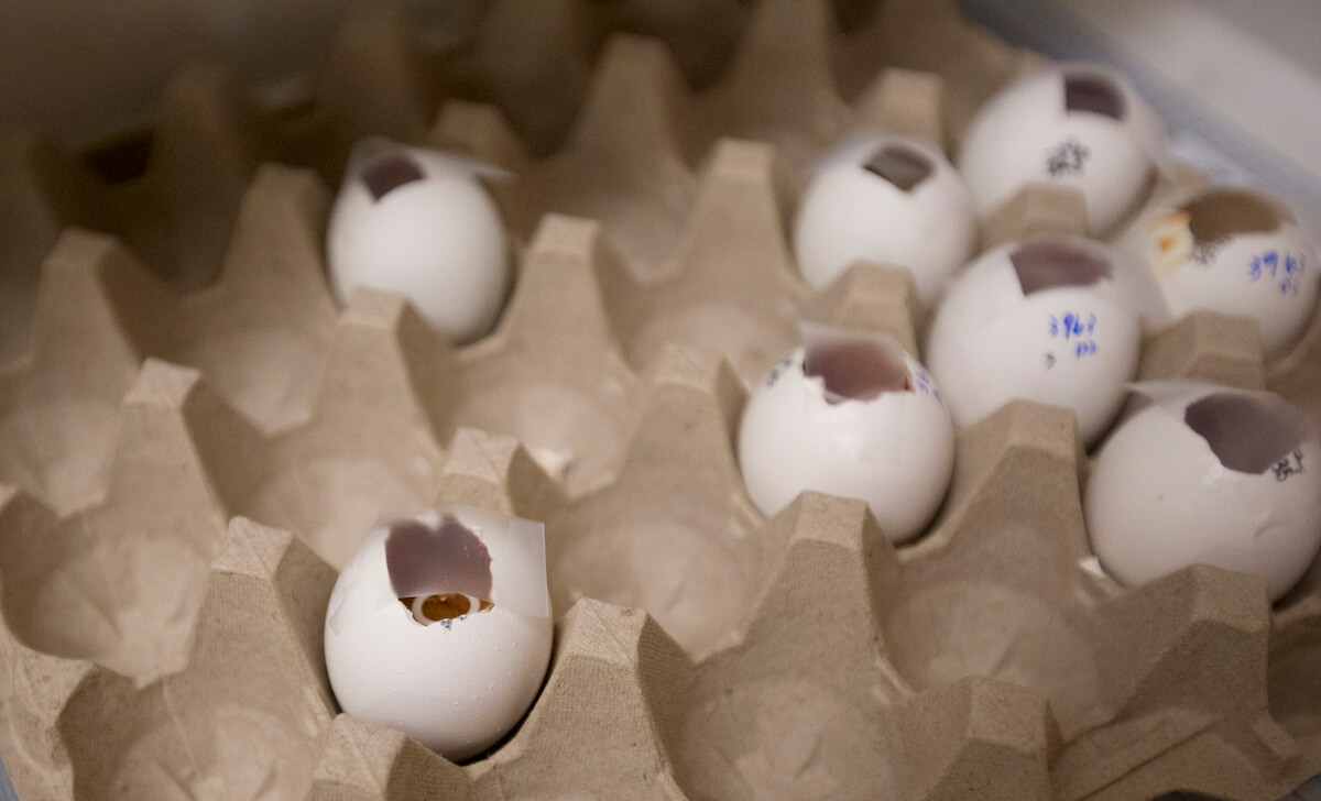
“Tumor Transplantation to the Chick Embryo”—a 1952 article published in Annals of the New York Academy of Sciences—noted the 1912 original research that “mouse and rat tumors would grow” when implanted in a chicken embryo’s chorio-allantoic membrane, known as CAM. The mid-century investigators confirmed that the approach showed promise because the embryo, lacking a defense against foreign tissue, would supply blood vessels and connective tissue for the tumors.
Advantages of eggs
Sikora aims to reinvent the use of eggs as a universal platform for cancer biology.
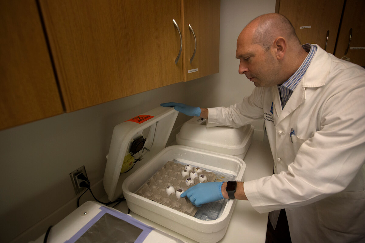
He is the academic director of Baylor’s Patient-Derived Xenograft and Advanced In Vivo Models Core, a part of Baylor’s research arm that has excelled at optimizing fertilized egg conditions and models. The core creates a collaborative environment for scientists to discuss potential studies using eggs.
“The eggs are so well vascularized that people used them as a platform to test angiogenesis,” said Sikora, referencing the process by which new blood vessels grow from pre-existing ones. “Does this compound induce blood vessels to grow? Can it grow cell lines to the primary tumor? We are pushing the system as far as we can to answer as many questions as possible.”
The CAM can support many human cancers, from the head and neck—Sikora’s areas of expertise—to breast, skin, ovarian, bladder and other cancer types. Even rare growths such as pancreatic neuroendocrine tumors can develop on the CAM, he said.
Sikora views fertilized eggs as “more faithful to the body” than a petri dish. In addition, the in vitro model enables quicker and more cost-effective pre-clinical trials of drugs because hundreds of samples can be tested at one time with results in about a week. Successful experiments can proceed to mice research.
Rowley’s interest sprang from the potential for efficiency. Mice experiments require more time and expense because of the need for special facilities and monitoring.
“It is a nice intermediate between doing an experiment on a cell culture dish and in a mouse,” he said.
Blocking prostate cancer
Rowley and Sikora worked together on research that involved metastasizing prostate cancer cells to a bovine bone. Bone metastasis is typically how prostate cancer kills.
Rebeca San Martin, Ph.D., the postdoctoral fellow who was working with Rowley at the time, wrote her thesis on the technology. The process involved placing a 5-millimeter bovine bone cube coded with tenascin-C—a protein often observed in cancerous tissue—on the CAM. She then placed an organoid made of cancer and stroma cells a few centimeters away to see if the cells would move to the bone. The cancer migrated, and those results were published in the November 2017 issue of Cancer Research.
Now, Rowley is investigating whether the cells traveled through the blood vasculature—a nod to one theory about how cancer cells metastasize to bone—and if a therapy targeting tenascin-C could neutralize the process.
“We think tenascin-C forms at the site of wound repairs,” he said. “Cancers are essentially wounds that don’t heal.”
Because the eggs offer financial and time-saving benefits for preliminary testing, Rowley believes they could be used to accelerate other experiments that otherwise would be conducted in mice.
“We got a lot of information from our study, but time will tell,” he said. “Could it be a better, cost-effective way to screen drugs? There may be other advantages or disadvantages to this method, but we don’t know yet.”